Cell membrane health
Cell Membrane Health: Why What Gets In and Out of Your Cells Determines Everything
By Lisa Ann
Most conversations about cellular health focus on what you put into your body — the quality of your food, the supplements you take, the nutrients you prioritize. These things matter enormously.
But there is a prior question that rarely gets asked: once those nutrients reach your cells, can the cells actually receive them?
The answer depends almost entirely on the health of a structure most people have never thought about: the cell membrane.
The cell membrane is not a passive container. It is one of the most sophisticated and metabolically active structures in biology — a dynamic, intelligent barrier that governs what enters each cell, what leaves it, how the cell communicates with its neighbors, and how it responds to the signals constantly arriving from hormones, neurotransmitters, immune molecules, and environmental inputs.
When cell membranes are healthy, the entire cellular communication network functions with precision. When they are compromised — built from the wrong materials, depleted of key structural components, or operating in a mineral-deficient environment — the downstream effects are wide-ranging and often completely misattributed.
Understanding membrane health is the second pillar of cellular resilience. And it explains a great deal about why people can be doing all the right things and still not getting the results they expect.
The Architecture of the Cell Membrane
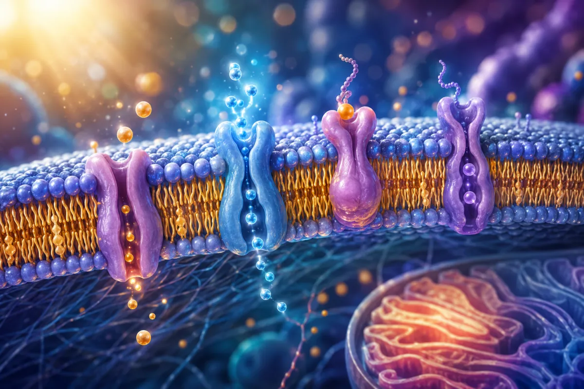
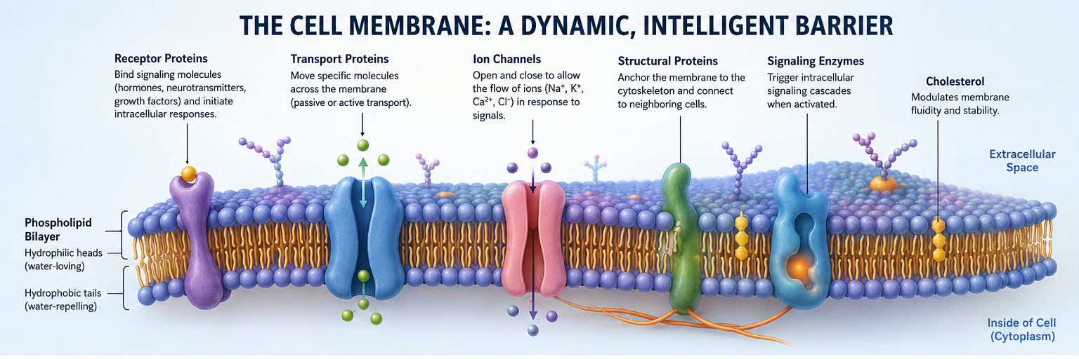
The cell membrane is a phospholipid bilayer — two sheets of phospholipid molecules arranged tail-to-tail, forming a continuous, fluid barrier around the cell. Each phospholipid molecule has a hydrophilic (water-attracting) head and two hydrophobic (water-repelling) fatty acid tails. This arrangement creates a structure that is simultaneously stable and fluid — firm enough to maintain cell integrity, flexible enough to allow the constant movement of proteins, channels, and receptors that cellular function requires.
Embedded within this bilayer is an extraordinary collection of functional proteins:
Receptor proteins that recognize and bind hormones, neurotransmitters, growth factors, and immune signaling molecules — translating extracellular signals into intracellular responses.
Transport proteins and pumps that move specific molecules across the membrane — either passively along concentration gradients, or actively against gradients using ATP.
Ion channels that open and close in response to electrical signals, chemical signals, or mechanical forces — allowing the controlled flow of sodium, potassium, calcium, and chloride ions that underlies all electrical activity in the body.
Structural proteins that connect the membrane to the cytoskeleton and to neighboring cells, maintaining tissue integrity.
Signaling enzymes that initiate intracellular cascades in response to receptor activation.
Cholesterol is also a critical structural component of the membrane, dispersed throughout the bilayer where it modulates fluidity — preventing the membrane from becoming too rigid in cold conditions or too fluid in warm ones.
This architecture is not static. The composition of the membrane — particularly the fatty acid profile of the phospholipids — changes over time in response to the fats consumed in the diet. The membrane is, in a meaningful sense, built from what you eat.
The Sodium-Potassium Pump: Your Body's Electrical Engine
Of all the proteins embedded in the cell membrane, none is more foundational to cellular function than the sodium-potassium ATPase pump — commonly called the sodium-potassium pump.
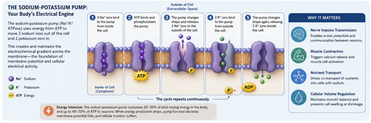
This remarkable protein runs continuously in virtually every cell in the body, doing one thing: moving three sodium ions out of the cell and two potassium ions in, for every molecule of ATP consumed. This seemingly simple exchange has profound consequences.
By maintaining higher concentrations of sodium outside the cell and potassium inside, the pump creates an electrochemical gradient across the membrane — a difference in both charge and ion concentration that stores potential energy the way a battery stores electrical charge. This gradient, called the membrane potential, is measured in millivolts and typically runs between -70 and -90 millivolts in a healthy resting cell.
Membrane potential is not incidental. It is the foundation of virtually all cellular electrical activity:
Nerve impulse transmission. Action potentials — the electrical signals that travel along neurons — are generated by the rapid, sequential opening of sodium and potassium channels along the nerve fiber. Without adequate membrane potential maintained by the sodium-potassium pump, neurons cannot fire reliably.
Muscle contraction. Every muscle contraction, from the beating of the heart to the blinking of an eye, depends on membrane potential changes triggering calcium release within muscle cells.
Nutrient transport. Many nutrients enter cells by co-transport — hitching a ride with sodium as it flows back into the cell along its concentration gradient. If the sodium gradient is inadequate, nutrient uptake suffers.
Cellular volume regulation. The pump helps maintain the osmotic balance that keeps cells from swelling or shrinking.
The sodium-potassium pump is extraordinarily energy-hungry. In the brain, it consumes an estimated 40 to 70 percent of total neuronal ATP. In the body as a whole, it accounts for roughly 20 to 30 percent of total resting energy expenditure.
This has a critical implication: when mitochondrial energy production is impaired (as discussed in the previous post in this series), the sodium-potassium pump is among the first systems to be compromised. Reduced ATP availability means reduced pumping activity, declining membrane potential, and a cascade of downstream cellular dysfunction.
The mitochondria and the membrane are not separate systems. They are profoundly interdependent.
What Determines Membrane Quality
If the membrane is built from what you eat, the quality of dietary fats becomes one of the most consequential nutritional variables in cellular health.
The Role of Omega-3 Fatty Acids
Omega-3 fatty acids — particularly DHA (docosahexaenoic acid) and EPA (eicosapentaenoic acid) — are among the most structurally important components of cell membranes. DHA in particular is highly concentrated in brain and neural tissue, where its unique molecular geometry contributes to the exceptional membrane fluidity required for rapid synaptic signaling.
Membranes rich in omega-3 fatty acids tend to be more fluid, more flexible, and more responsive. Receptor proteins embedded in omega-3-rich membranes can move more freely to bind their ligands, increasing receptor sensitivity. Ion channels open and close more efficiently. Signaling cascades initiate more readily.
The ratio of omega-6 to omega-3 fatty acids in the membrane is particularly important. The modern diet, which is heavily skewed toward omega-6 fats from seed oils and processed foods, tends to produce membranes with a much higher omega-6 content than what human biology evolved with. While omega-6 fats are not inherently harmful, excess omega-6 relative to omega-3 contributes to more pro-inflammatory signaling at the membrane level and reduced receptor sensitivity.
The Problem with Damaged and Oxidized Fats
Beyond the omega-6 to omega-3 ratio, the integrity of the fats incorporated into membranes matters enormously. Fats are chemically fragile — they oxidize readily when exposed to heat, light, and oxygen. Repeatedly heated cooking oils, processed foods containing partially hydrogenated fats, and foods fried at high temperatures all contain oxidized and structurally altered fatty acids.
When these damaged fats are incorporated into cell membranes instead of intact, undamaged ones, the consequences are significant:
The membrane becomes less fluid and less responsive. Receptors do not sit correctly in the bilayer. Channels do not open and close with normal efficiency. The signaling environment becomes noisy and imprecise.
This is one of the mechanisms by which a diet high in processed and industrially produced fats — even in the absence of obvious caloric excess — can impair cellular function in ways that generate symptoms without necessarily showing up on standard laboratory tests.
Phospholipids and Membrane Repair
Phospholipids are not just the structural backbone of the membrane — they are also actively involved in cell signaling. Phosphatidylcholine, phosphatidylserine, and phosphatidylinositol each have specific functional roles beyond structure, and their adequate availability supports both membrane integrity and the signaling cascades that depend on membrane-derived molecules.
Phosphatidylserine, for example, is concentrated in neuronal membranes and plays a role in neurotransmitter release, neuroplasticity, and the regulation of the stress response through its effects on cortisol signaling. Its depletion in aging and under chronic stress is one reason membrane-targeted nutrition is so relevant to cognitive and neurological health.
Calcium Ion Channels and Why Their Regulation Matters
Embedded in the cell membrane are voltage-gated calcium channels (VGCCs) — proteins that open in response to changes in membrane voltage, allowing calcium ions to flow rapidly into the cell. This calcium influx triggers an extraordinary range of downstream biological events: neurotransmitter release at synapses, muscle contraction, enzyme activation, gene expression, and cell division.
Calcium signaling must be exquisitely precise. Inside a resting cell, calcium concentrations are kept extremely low — roughly 10,000 times lower than outside the cell. This massive gradient means that when calcium channels open, even briefly, a large amount of calcium flows in relative to the baseline intracellular concentration. The signal is sharp, clear, and rapidly terminated as calcium is pumped back out of the cell.
This precision is what makes calcium such an effective signaling molecule. The same property also makes dysregulation of calcium channels particularly consequential.
When voltage-gated calcium channels open too frequently, stay open too long, or become hypersensitive to activating signals, calcium accumulates inside the cell beyond normal signaling levels. The consequences of sustained intracellular calcium elevation include:
Chronic activation of calcium-dependent inflammatory pathways
Continuous excitation of neurons, contributing to hyperexcitability and anxiety
Impaired muscle relaxation, producing chronic tension
Mitochondrial dysfunction, as excess intracellular calcium disrupts mitochondrial membrane potential
Disrupted sleep architecture, as calcium-dependent neurotransmitter systems are persistently activated
Long-term excitotoxic effects in neural tissue at high levels
What causes voltage-gated calcium channels to become dysregulated? Several factors are well-documented. Magnesium deficiency is among the most important — magnesium acts as a natural physiological blocker of VGCCs, sitting in the channel and preventing excessive calcium entry. When magnesium is depleted, the natural brake on calcium channel activity is removed.
Electromagnetic frequency (EMF) exposure is another documented activator of VGCCs — a finding with significant implications that I address in detail in the EMF installment of this series. And chronic stress, which alters membrane potential and increases sympathetic nervous system tone, also contributes to calcium channel dysregulation through multiple pathways.
Receptor Sites and the Problem of Diminished Sensitivity
The cell membrane is studded with receptor proteins — each designed to recognize a specific signaling molecule and initiate a specific cellular response. The sensitivity and density of these receptors determines how effectively the cell can respond to incoming signals.
Insulin receptors govern glucose uptake and metabolic function. Hormone receptors translate estrogen, progesterone, testosterone, cortisol, and thyroid hormone signals into cellular action. Neurotransmitter receptors govern mood, motivation, sleep, pain perception, and cognitive function. Immune receptors coordinate the inflammatory and anti-inflammatory responses.
When receptor sensitivity is reduced — whether from membrane lipid quality issues, chronic overstimulation of receptors by excess circulating ligands, or physical displacement of receptor proteins by structural membrane changes — the cell becomes less responsive to normal physiological signals.
This receptor desensitization is the mechanism behind several of the most common patterns seen in functional health practice:
Insulin resistance is fundamentally a problem of impaired insulin receptor signaling — the pancreas produces insulin, but the cellular response is blunted. The connection between membrane lipid quality and insulin receptor function has been documented in research linking omega-3 fatty acid incorporation into membranes with improved insulin sensitivity.
Thyroid resistance — normal or even elevated thyroid hormone levels on blood tests, with symptoms consistent with hypothyroidism — often reflects impaired thyroid hormone receptor signaling at the cellular level rather than inadequate hormone production.
Hormone imbalance symptoms in the presence of normal hormone levels may indicate receptor sensitivity issues rather than production deficiencies — a distinction that fundamentally changes the appropriate intervention.
Neurotransmitter dysregulation — including the mood, motivation, anxiety, and sleep disturbances associated with imbalances in serotonin, dopamine, and GABA — is not only a function of neurotransmitter production but also of receptor density and sensitivity in the membranes of target neurons.
This is one of the reasons that addressing membrane health — through targeted nutrition, mineral restoration, and reduction of membrane-damaging inputs — often produces improvements in hormone signaling, mood, metabolism, and cognitive function that cannot be explained by changes in hormone or neurotransmitter levels alone.
The Mineral-Membrane Connection
Membrane function and mineral status are inseparable. The sodium-potassium pump requires magnesium as a cofactor to function. Calcium channel regulation depends on magnesium as a natural blocker. The membrane potential itself is determined by the concentration gradients of sodium, potassium, calcium, and chloride across the membrane — gradients that are maintained by mineral-dependent pumps and transporters.
When mineral status is depleted — as is extremely common under chronic stress, with poor dietary diversity, or with gut absorption challenges — the electrical environment of the membrane degrades. Membrane potential declines. Ion channel function becomes less precise. The sodium-potassium pump runs less efficiently, consuming more ATP for less pumping activity.
The Hair Tissue Mineral Analysis (HTMA) is one of the most informative assessments I use to understand the mineral environment affecting membrane function. By measuring mineral patterns in tissue rather than serum, the HTMA provides a window into the mineral dynamics that influence membrane potential, stress physiology, and electrolyte balance over a two-to-three month window — revealing patterns that standard blood panels routinely miss.
Why Healthy Inputs Don't Always Produce Healthy Outcomes
This is the question that brings many people to functional health practice after years of doing things conventionally right: I am eating well, supplementing thoughtfully, exercising, managing stress — why don't I feel better?
The answer often lies in what we have been discussing throughout this post. When cell membranes are built from suboptimal materials, when the sodium-potassium pump is running at reduced capacity due to mineral depletion, when receptor sensitivity has declined, when calcium channels are dysregulated — the cellular machinery that is supposed to receive, process, and respond to all those good inputs is simply not functioning well enough to translate them into the outcomes you are working toward.
Supplements do not bypass the membrane. Hormones cannot signal effectively through a degraded receptor environment. Nutrients transported by sodium co-transport systems are impaired when the sodium gradient is inadequate. The membrane is not a detail. It is the interface through which everything else works.
This is why membrane health is not an advanced or optional topic in cellular nutrition. It is foundational — upstream of nearly everything else, and often the missing piece that explains why intelligent, committed people plateau despite doing so much right.
Supporting Membrane Health Practically
Membrane restoration is not a rapid process — the phospholipid composition of membranes turns over gradually, reflecting months of dietary fat intake. But the changes that come from sustained attention to membrane health tend to be durable and wide-ranging.
The most important practical priorities:
Prioritize high-quality omega-3 sources. Cold-water fatty fish (salmon, sardines, mackerel, herring), grass-fed and pasture-raised animal products, and high-quality fish or algae oil supplements all increase the DHA and EPA content of cell membranes. This is the single most impactful dietary change for membrane fluidity and receptor sensitivity.
Reduce damaged and oxidized fats. Minimizing industrial seed oils (particularly those used at high heat), processed foods, and fried foods reduces the incorporation of oxidized fatty acids into membrane structure.
Support phospholipid availability. Egg yolks, liver, and lecithin are rich in phosphatidylcholine and other membrane phospholipids. Targeted supplementation with phosphatidylserine can be particularly relevant for neurological and adrenal support.
Ensure fat-soluble vitamin adequacy. Vitamins A, D, E, and K all play roles in membrane function — vitamin E as a fat-soluble antioxidant that protects membrane lipids from oxidation, vitamins A and D in the regulation of gene expression that influences membrane protein production.
Restore mineral balance. Magnesium is the priority — its role in sodium-potassium pump function, VGCC regulation, and ATP production makes it central to membrane electrical health. Potassium and sodium balance matter as well. HTMA assessment is the most precise way to understand an individual's mineral status and guide targeted restoration.
Address gut integrity. The absorption of fat-soluble nutrients, essential fatty acids, and fat-soluble vitamins depends on healthy intestinal fat absorption — bile production, pancreatic enzyme function, and intestinal membrane integrity all influence how effectively the building blocks of healthy cell membranes actually reach the bloodstream.
The Bigger Picture
The cell membrane is where the outside world meets the inside of the cell. Every signal, every nutrient, every hormone, every environmental input that influences cellular behavior does so through the membrane — through its channels, its receptors, its pumps, and its selective permeability.
When the membrane is healthy, the cell is in genuine conversation with its environment. When it is compromised, that conversation becomes distorted — signals are missed, nutrients are blocked, electrical function degrades, and the entire cellular communication network loses precision.
Building membrane health is not about a single nutrient or a single intervention. It is about creating and sustaining the conditions in which the membrane can maintain its integrity, its fluidity, and its extraordinary functional capacity.
When that happens, the downstream effects — on energy, on hormonal signaling, on cognitive function, on mood, on recovery, on resilience — tend to be among the most comprehensive and durable improvements people experience in their health.
If you are curious about your mineral status and how it may be affecting membrane function, the HTMA assessment provides a detailed picture of the mineral patterns that underlie cellular electrical health. The NeuroLongevity Quickstart includes a Cellular Resilience Test that looks more broadly at what may be interfering with cellular function. [Learn more here]


